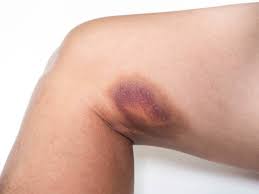
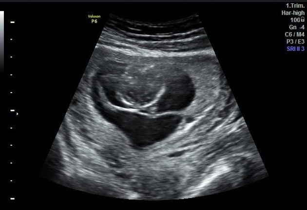
Hematomas are the accumulation of blood outside of blood vessels. This can occur in various parts of the body. Hematomas can cause pain and discomfort, but in some cases drainage of the hematoma will become necessary to reduce the symptoms and encourage proper healing. So, when exactly does a hematoma need to be drained? Let’s take a look into when and why hematoma drainage may be necessary.
Understanding hematomas
Firstly it is important to have a clear understanding of what hematomas are and how they form. A hematoma can look like a bruise, but bruises occur due to damage to small blood vessels rather than large ones. Hematomas occur in larger blood vessels, including veins, and arteries. A hematoma is essentially a collection of blood that has leaked from damaged blood vessels, pooling in the injured area. This can occur as a result of trauma, surgery, or underlying medical conditions that affect blood clotting.
Hematomas can develop in various parts of the body, including soft tissues, muscles and organs, inside the skull, on the scalp, ears, septum, bones, fingernails and toenails (subungual hematoma). The severity of a hematoma can vary, ranging from mild discomfort to significant pain and swelling. In some cases, the accumulation of blood can slow down healing and the recovery process.
Common indications for hematoma drainage
Size
One of the main considerations for hematoma drainage is the size of the haematoma. Large hematomas, particularly those that continue to expand over time, can exert pressure on surrounding tissues, block blood flow, and cause considerable pain. In such cases, drainage becomes necessary to relieve the pressure and prevent further complications.
Persistent pain and discomfort
When hematomas occur in sensitive areas or put pressure on nerves, they are often accompanied by pain and discomfort. If conservative measures such as pain medication and rest fail to alleviate the pain, drainage may be required to provide relief and promote healing.
Impaired functionality
Hematomas can lead to functional impairment, restricting movement or causing disorders in the affected area. A large hematoma in the joint may limit the range of motion and hinder mobility. In such situations, drainage can help restore functionality and facilitate rehabilitation.
Risk of infection
Hematomas that are at risk of becoming infected may also require drainage. When blood pools outside blood vessels, it creates an environment conducive to bacterial growth. If there are signs of infection such as redness, warmth, or fever, drainage may be necessary to prevent the spread of infection and promote healing.
Underlying medical conditions
Individuals with certain medical conditions, such as coagulation disorders or compromised immune systems, may be more susceptible to complications from hematomas. In such cases, drainage may be recommended even for smaller hematomas, to prevent potential complications and promote optimal healing.
When considering hematoma drainage, healthcare professionals must take into consideration the overall health, medical history, and individual preferences of patients.
The hematoma drainage procedure
When the decision to drain a hematoma is made, the procedure typically involves the insertion of a catheter or needle to remove the accumulated blood. The exact technique may vary depending on the size, location and type of hematoma. Here are some common methods used for hematoma drainage:
Percutaneous drainage
Percutaneous drainage involves the insertion of a catheter or needle directly into the hematoma under imaging guidance. This allows for precise placement and effective drainage of the accumulated blood. The catheter can be left in place for a specific duration to ensure complete drainage and prevent re-accumulation.
Surgical drainage
Surgical drainage may be necessary, especially for larger or more complex hematomas. This procedure involves making an opening to access the hematoma to drain the blood manually. Surgical drainage may be preferred when other interventions have been unsuccessful.
Ultrasound-guided drainage
Ultrasound guidance can be carried out to secure the accurate placement of the drainage catheter or needle. This technique allows for real-time visualization of the hematoma and surrounding structures, reducing the risk of complications and improving the success rate of the drainage procedure.
Complications
While hematoma drainage is generally a safe and effective procedure, some potential complications and considerations need to be taken into account. These include:




















Infection
Any invasive procedure carries a risk of infection. Proper sterile techniques and vigilant monitoring are crucial to minimize the risk of introducing bacteria into the hematoma or surrounding tissues during the drainage procedure.
Bleeding
There is a potential risk of bleeding during or after hematoma drainage. This risk is higher in individuals with underlying bleeding disorders or those taking blood thinners such as warfarin.
Recurrence
In some cases, hematomas may re-accumulate after drainage. This can be due to ongoing bleeding, inadequate drainage, or the persistence of underlying conditions. Close follow-up and appropriate management strategies are necessary to prevent recurrence and ensure optimal healing.
Fistula formation
In rare instances, drainage procedures can lead to the formation of abnormal connections between the hematoma and adjacent structures, such as the bowel, pancreas, or biliary system. These fistulas can cause further complications and may require additional interventions for resolution.
Conclusion
Hematoma drainage is useful in cases where the accumulation of blood causes significant discomfort with symptoms, loss of movement, or poses a risk of infection. The decision to drain a hematoma is based on various factors, including size, symptoms, and associated risks. While hematoma drainage is rarely life-threatening, it is essential to consider potential complications and seek medical care if needed. Healthcare providers can determine when hematoma drainage is necessary and provide medical advice.
Sources
About NowPatient's Editorial Process
- Written by clinical authors
- Reviewed every 2 years
- Conflicts of interest declared
NowPatient's medical content is selected by a panel of practising clinicians (pharmacists, doctors and allied healthcare professionals) and researched by an in-house team of writers who all hold scientific or clinical backgrounds. Sponsorship and conflicts of interest are openly declared.
Every article is peer-reviewed by a clinician for clinical accuracy, evidence base and accessibility, including consideration of the particular characteristics set out in the UK Equality Act 2010. Only then is it released for publication against fixed placement guidelines.
All content is cycle reviewed (typically every two years) and updated sooner if evidence, guidelines or a complaint require it. Changes follow a formal change control procedure and are reflected in the "last updated" date on each article. NowPatient is governed by MiHealth; UK pharmacy services are provided by MiHealth Pharmacy (GPhC 1036487).